Back
BackWhat is Histology?
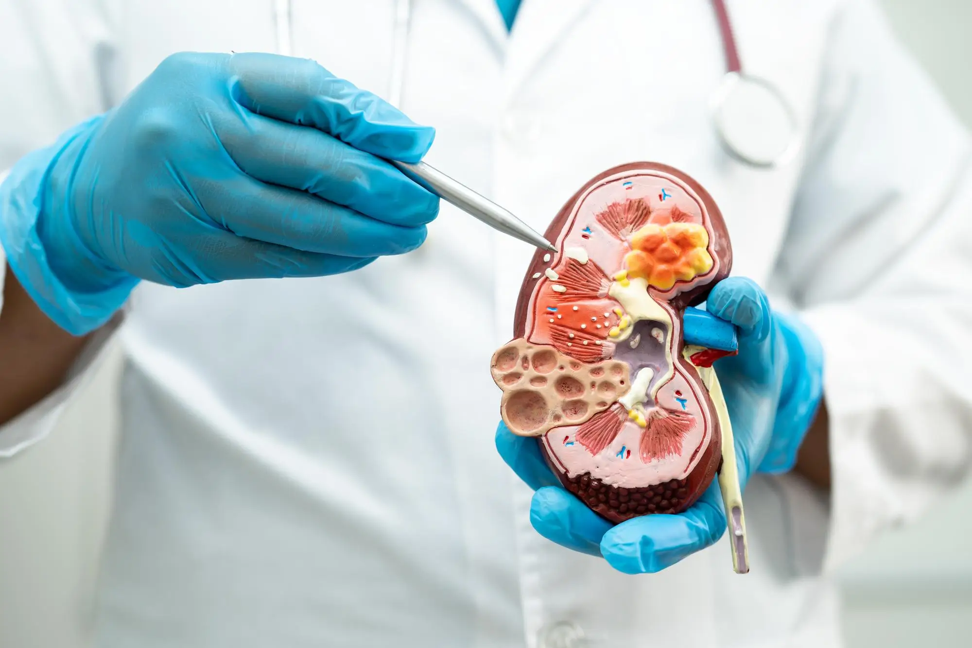
Histology is the study of body tissues at a microscopic level. It provides insights into the structure of cells and organs. Histological analysis helps to identify cellular alterations that might signal different diseases, making histology an indispensable diagnostic tool.
About usOur goal is to ensure the best possible medical care, covering the entire spectrum of advanced clinical pathology diagnostics.
OLYMP CDL provides an extensive range of morphological examinations for healthcare, dental, outpatient and hospital institutions.
Why does the choice turn to us?

Efficient Logistics
Streamlined transportation routes ensure prompt delivery

Digital Services
Innovative applications for submitting requests and receiving reports

Constant Engagement
Participation in scientific and practical conferences and close with clinicians
We are proud of the strong satisfaction rates among our partners and doctors, achieved through our reliability and clinical engagement.
Our TeamJoint efforts for the good of your health!
We believe that teamwork is the key to success. In our laboratory, each specialist focuses on their area of expertise and passion to achieve the best possible results.
Our advantages
1
Synergy of Expertise
By combining our knowledge and ideas, we deliver services at the highest level of modern science.
2
Technology and Innovation
Cutting-edge equipment and collaboration with a global network of scientists, doctors, and specialists unlock endless possibilities for us.

Pathological Anatomy include the following components:
- Histopathology – the study of tissues to detect microscopic changes
- Cytopathology — the study of cells detached from tissues or present in bodily fluids
At OLYMP CDL biological material is processed using advanced technical and methodological solutions. This enables us to:
- Arrive at accurate diagnosis based on histological and cytological data
- Provide a foundation for further therapeutic or diagnostic decisions in patient treatment
We employ all possible methods to ensure diagnostic accuracy and successful treatment.
Additional advanced technologies can be applied when needed:
- Histochemistry and Cytochemistry
- Immunohistology and Immunocytology
- Molecular Pathology
- Mass Spectroscopy
Pathology (derives from the Greek roots πάθος, meaning ‘suffering’, and – λόγος, meaning ‘study of’) is a field of study focused on understanding the nature of diseases and health. Despite common misconceptions, pathology is not associated with forensic medicine.
Pathology today — is a field devoted to analyzing disease-induced changes in the structure and function of human cells, tissues and organs.
The Role of a Pathologist
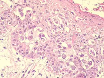
One of the key responsibilities of a pathologist is detecting malignant tumors.
- Physicians send tissue or cell samples obtained through biopsy or surgery for examination.
- Only a pathologist can accurately classify a tumor, something that cannot be achieved with other laboratory methods.
Diagnosis and Treatment
A diagnosis based on additional studies (such as immunohistochemistry or molecular pathology) serves as the foundation for determining the treatment strategy:
- Surgical intervention
- Chemotherapy
- Radiation therapy
Pathology today is the cornerstone of medicine, providing accurate diagnostics and a personalized approach to treatment.
Trust. Openness. Friendliness.
We build our work on the principles of trustworthy, open, and friendly collaboration with medical organizations and colleagues. Our team adheres to an approach based on:
- Teamwork
- Responsibility and responsiveness
- Respectful communication
Each specialist in our laboratory is evaluated and developed according to their individual skills and area of responsibility.
To achieve the highest level of diagnostics in histopathology, cytology, and molecular pathology, we rely on the following:
Mutual support
Employees work closely together, assisting one another.Continuous development
Regular training for both medical and non-medical staff.
By combining professionalism, advanced equipment and a cohesive team, we ensure high-quality diagnostics at every stage.
Proper Delivery = Fast Results
To ensure accurate and rapid results, it is essential to follow strict requirements for the preparation and transportation of biological samples.
Material Fixation
Biopsy samples and small tissue specimens should be fixed in 10% formalin (ratio of at least 1:10).
Processing of Large Samples:
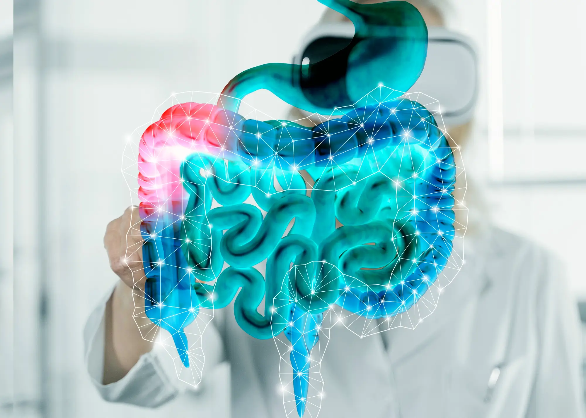
- Organs (e.g., intestines, stomach) should be opened, cleaned, and fixed in formalin
- For large samples (liver, kidneys, spleen), deep incisions are recommended to allow formalin penetration without compromising tissue structure
Requirements for Examined Material
Tumor Samples:
- Should contain the highest possible number of tumor cells
- Samples with necrosis or hemorrhage are of limited diagnostic value
Formalin Fixation:
- Mandatory to prevent DNA degradation
- Acidic decalcification renders the material unsuitable for molecular studies
- EDTA decalcification is possible but reduces analysis quality
Temperature Conditions
High temperatures and vacuum negatively impact DNA quality.
Indications for Microsatellite Analysis
Microsatellite analysis is performed in the following cases:
- Colorectal cancer before the age of 50
- Synchronous or metachronous carcinomas associated with HNPCC-related tumors (colon, rectum, stomach, ovaries, etc.)
- MSI-histology:
- Tumor-infiltrating lymphocytes
- Tumor-infiltrating lymphocytes
- Medullary growth
- Family history:
- First-degree relative with colorectal cancer before the age of 50
- At least two relatives with colorectal cancer or HNPCC-related tumors
According to the Ministry of Health of the Republic of Kazakhstan, the following receptor expressions must be determined for each breast carcinoma:
Ductal Carcinoma In Situ (DCIS):
- Estrogen Receptor
- Progesterone Receptor
Invasive Carcinomas:
- Estrogen Receptor (ER)
- Рецептор прогестерона (PR)
- Her2
Receptor expression is of two key importance:
Individual Risk Assessment:
- The absence of ER and/or PR expression corresponds to a worse prognosis.
- HER2 overexpression also indicates an unfavourable prognosis.
Basis for Therapy:
- Adjuvant chemotherapy (Tamoxifen, Aromatase inhibitors: Letrozole, Anastrozole, Exemestane) is used for PR/ER-positive tumors.
- Herceptin therapy is indicated in cases of HER2 overexpression.
Receptor expression is assessed semi-quantitatively.
It is crucial to consider that fixation artifacts in tissue samples may cause regional receptor expression. Therefore, if neoadjuvant therapy is not prescribed, receptor expression analysis should be performed on resected tissue material.
Standards for Receptor Evaluation
Number of Positive Cells
| Number of Positive Nuclei | Score |
|---|
| Not detected | 0 |
| Less than 10% | 1 |
| 10–50% | 2 |
| 51–80% | 3 |
| More than 80% | 4 |
Интенсивность окрашивания
| Intensity | Score |
|---|
| No staining | 0 |
| Low | 1 |
| Moderate | 2 |
| Strong | 3 |